X-Ray vs.
MRI
With spinal imaging, your physician can pinpoint the area and severity of
your problem prior to beginning IDD Spinal Decompression Therapy. This also helps to determine if you are a
candidate for Spinal Decompression.
If you have any type of neck or back disorder and your physician does not
refer you for MRI imaging studies prior to beginning any type of spinal decompression program, this will only
result in guess work as to which spinal segment and disc to treat.
The physician will also have a lack of knowledge if there are any cautions
or contraindications as to why you should not have spinal decompression therapy which can result in
aggravating or causing additional injuries to your spine.
MRI and CT scans can be used to better evaluate the "soft tissues" such as
the nerves, discs and ligaments. When there is a disc herniation, X-rays may look completely normal whereas
the MRI can show the extent of nerve compression caused by the disc herniation.
I have included some samples below so that you may understand the
importance of proper imgaing studies prior to beginning any spinal decompression
treatment.
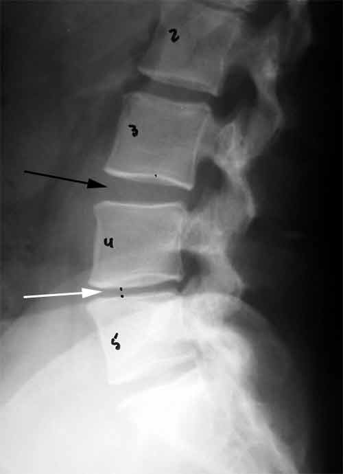
 The X-ray to the left is that of the lumbar [low back] spine. The vertebrae
are number from 2-5. The disc spaces are in-between the vertebrae. The X-ray to the left is that of the lumbar [low back] spine. The vertebrae
are number from 2-5. The disc spaces are in-between the vertebrae.
Each disc space is associated with 2 vertebrae and, therefore, has two numbers for its name. For example, the white
arrow points to the L4/5 disc space.
Note the difference between the L3/4 [black arrow] and L4/5 [white arrow] disc spaces.
The white arrow shows the collapse, "degenerative" disc compared to the "normal" L3/4 disc
space.
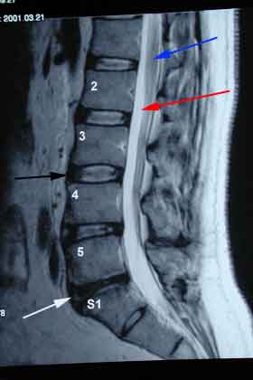
 The MRI to
the left is an image of the The MRI to
the left is an image of the
lumbar spine [low
back]. The vertebrae are marked with numbers; one can see lumbar vertebrae 2 through 5 and finally the S1 vertebra
[which is the Sacral #1 vertebrae].
The discs are in-between the vertebrae are number accordingly.
For example, the L3/4 disc has a black arrow pointing to it. The discs always have
2 numbers for identification.
The Red
arrow points to the fluid in spinal canal; this fluid appears as a whitish color on
the MRI.
The Blue arrow points to a nerve
roots in the canal.
When you look closely at the discs, you notice that the L5/S1 disc
[white arrow] is dark compared to the rest of the discs
which are more whitish in color. The dark disc is considered degenerated and has lost its "normal
fluid".
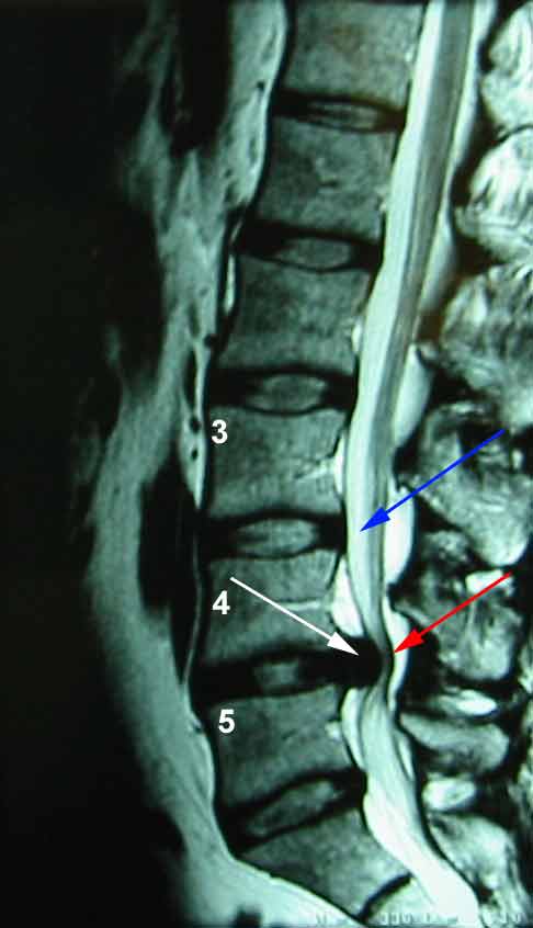
 The MRI to the left shows a similar view
of the low back. The spinal canal with the whitish fluid is violated by a large, golf sized disc herniation as
marked with the white arrow. The MRI to the left shows a similar view
of the low back. The spinal canal with the whitish fluid is violated by a large, golf sized disc herniation as
marked with the white arrow.
The Red arrow points to the
spinal nerves which are being pushed back by the disc herniation.
The Blue arrow points to the
white spinal fluid.
| 


